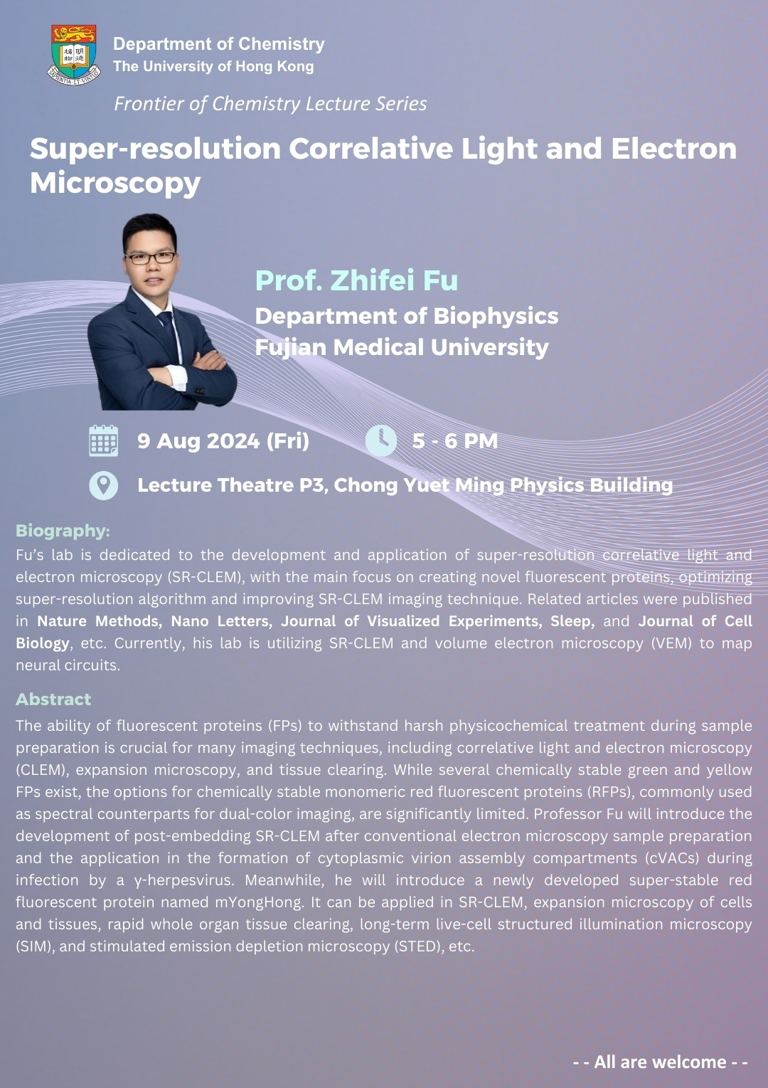
| Date | 09 Aug 2024 |
| Time | 5:00 pm - 6:00 pm (HKT) |
| Venue | Lecture Theatre P3, Chong Yuet Ming Physics Building |
| Speaker | Prof. Zhifei Fu |
| Institution | Department of Biophysics, Fujian Medical University |

Title:
Super-resolution Correlative Light and Electron Microscopy
Schedule:
Date: 9th August, 2024 (Friday)
Time: 5 - 6 pm (HKT)
Venue: Lecture Theatre P3, Chong Yuet Ming Physics Building
Speaker:
Prof. Zhifei Fu
Department of Biophysics
Fujian Medical University
Biography:
Fu’s lab is dedicated to the development and application of super-resolution correlative light and electron microscopy (SR-CLEM), with the main focus on creating novel fluorescent proteins, optimizing super-resolution algorithm and improving SR-CLEM imaging technique. Related articles were published in Nature Methods, Nano Letters, Journal of Visualized Experiments, Sleep, and Journal of Cell Biology, etc. Currently, his lab is utilizing SR-CLEM and volume electron microscopy (VEM) to map neural circuits.
Abstract:
The ability of fluorescent proteins (FPs) to withstand harsh physicochemical treatment during sample preparation is crucial for many imaging techniques, including correlative light and electron microscopy (CLEM), expansion microscopy, and tissue clearing. While several chemically stable green and yellow FPs exist, the options for chemically stable monomeric red fluorescent proteins (RFPs), commonly used as spectral counterparts for dual-color imaging, are significantly limited. Professor Fu will introduce the development of post-embedding SR-CLEM after conventional electron microscopy sample preparation and the application in the formation of cytoplasmic virion assembly compartments (cVACs) during infection by a γ-herpesvirus. Meanwhile, he will introduce a newly developed super-stable red fluorescent protein named mYongHong. It can be applied in SR-CLEM, expansion microscopy of cells and tissues, rapid whole organ tissue clearing, long-term live-cell structured illumination microscopy (SIM), and stimulated emission depletion microscopy (STED), etc.
- - ALL ARE WELCOME --
