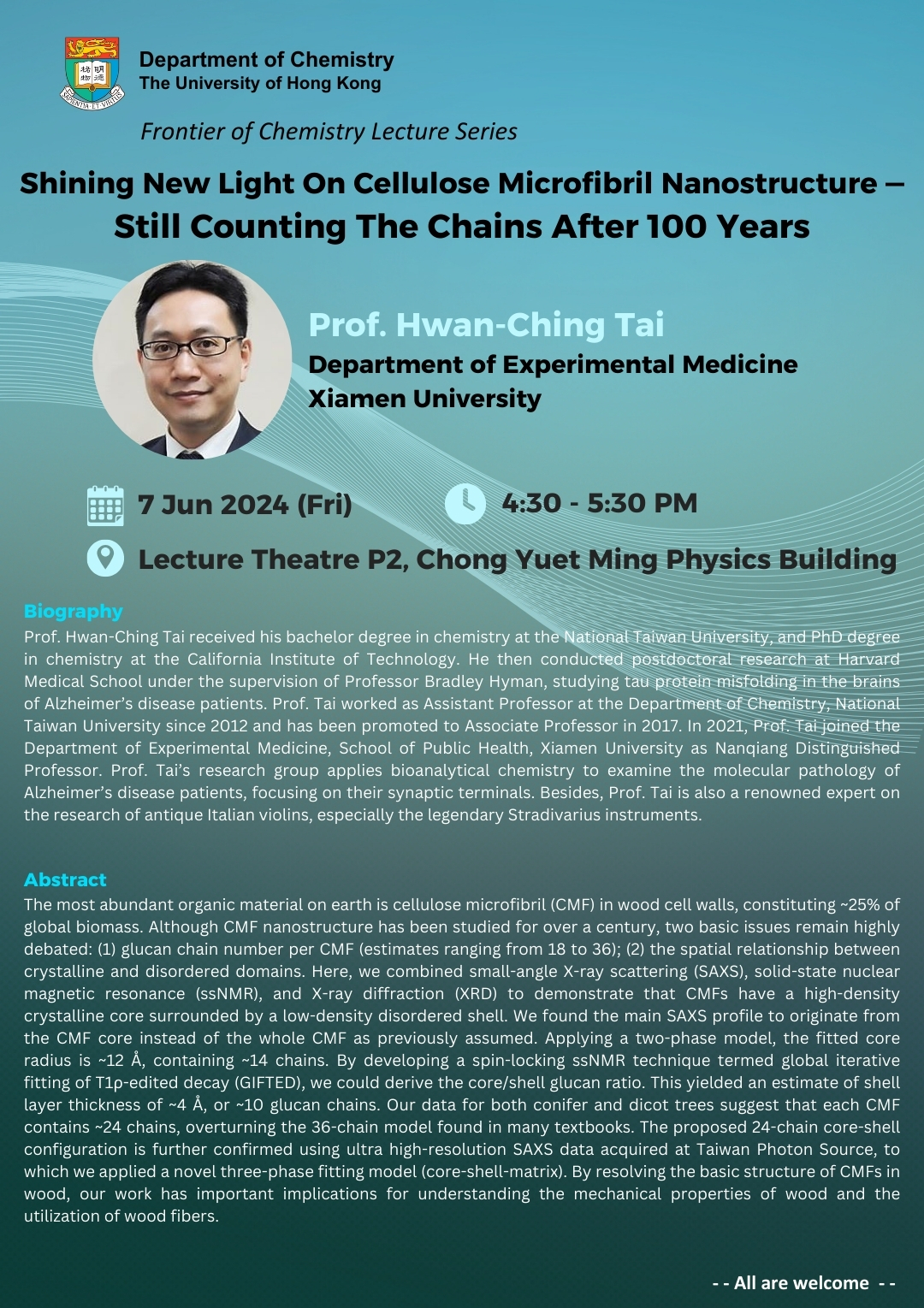
| Date | 07 Jun 2024 |
| Time | 4:30 pm - 5:30 pm (HKT) |
| Venue | Lecture Theatre P2, Chong Yuet Ming Physics Building |
| Speaker | Prof. Hwan-Ching Tai |
| Institution | Department of Experimental Medicine, Xiamen University |

Title:
Shining New Light On Cellulose Microfibril Nanostructure — Still Counting The Chains After 100 Years
Schedule:
Date: 7 Jun, 2024 (Friday)
Time: 4:30 - 5:30 pm (HKT)
Venue: Lecture Theatre P2, Chong Yuet Ming Physics Building
Speaker:
Prof. Hwan-Ching Tai
Department of Experimental Medicine
Xiamen University
Biography:
Prof. Hwan-Ching Tai received his bachelor degree in chemistry at the National Taiwan University, and PhD degree in chemistry at the California Institute of Technology. He then conducted postdoctoral research at Harvard Medical School under the supervision of Professor Bradley Hyman, studying tau protein misfolding in the brains of Alzheimer’s disease patients. Prof. Tai worked as Assistant Professor at the Department of Chemistry, National Taiwan University since 2012 and has been promoted to Associate Professor in 2017. In 2021, Prof. Tai joined the Department of Experimental Medicine, School of Public Health, Xiamen University as Nanqiang Distinguished Professor. Prof. Tai’s research group applies bioanalytical chemistry to examine the molecular pathology of Alzheimer’s disease patients, focusing on their synaptic terminals. Besides, Prof. Tai is also a renowned expert on the research of antique Italian violins, especially the legendary Stradivarius instruments.
Abstract:
The most abundant organic material on earth is cellulose microfibril (CMF) in wood cell walls, constituting ~25% of global biomass. Although CMF nanostructure has been studied for over a century, two basic issues remain highly debated: (1) glucan chain number per CMF (estimates ranging from 18 to 36); (2) the spatial relationship between crystalline and disordered domains. Here, we combined small-angle X-ray scattering (SAXS), solid-state nuclear magnetic resonance (ssNMR), and X-ray diffraction (XRD) to demonstrate that CMFs have a high-density crystalline core surrounded by a low-density disordered shell. We found the main SAXS profile to originate from the CMF core instead of the whole CMF as previously assumed. Applying a two-phase model, the fitted core radius is ~12 Å, containing ~14 chains. By developing a spin-locking ssNMR technique termed global iterative fitting of T1ρ-edited decay (GIFTED), we could derive the core/shell glucan ratio. This yielded an estimate of shell layer thickness of ~4 Å, or ~10 glucan chains. Our data for both conifer and dicot trees suggest that each CMF contains ~24 chains, overturning the 36-chain model found in many textbooks. The proposed 24-chain core-shell configuration is further confirmed using ultra high-resolution SAXS data acquired at Taiwan Photon Source, to which we applied a novel three-phase fitting model (core-shell-matrix). By resolving the basic structure of CMFs in wood, our work has important implications for understanding the mechanical properties of wood and the utilization of wood fibers.
- ALL ARE WELCOME -
